Channel treatment under the microscope
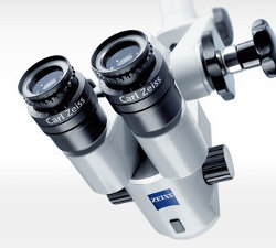
Root canal treatment under a microscope, thanks to the large enlargement of the tooth, allows the doctor to develop and fill the root canals much more accurately. Properly performed duct treatment is designed to remove the infected contents of the root canals, and then fill them up to the top of the root. Our office often reports patients with toothache, which were previously treated canalally. The causes of such pain can be many:
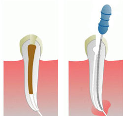
- Underfilling of root canals (Fig. 1). The cause of pain is bacteria developing in an unfilled episode, which form inflammation visible in the photo as a darker place around the root. Dental microscope allows for unprecedented visibility of the treatment field allowing the doctor to properly cure.
- Leaving the channel unfound (fig. 2). As a result of such action in the channel requiring filling, bacteria develop, preventing the bones from healing around the root, thereby causing pain to the patient. Treatment under a microscope allows you to enlarge the treatment area up to 30X, so finding all channels does not make it difficult.
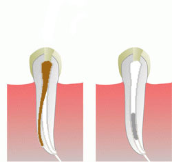
- A difficult to diagnose cause of pain is the filling of only one branch of the canal (Fig. 3). Working without a microscope, the doctor is not able to see the branching of the canal, which is located, for example, in the middle of its course. In such a situation, it occurs either to develop its channels only to branch, or to develop and fill only one branch.
- Fracture of the tool in the root canal (Fig. 4), is a common complication of endodontic treatment. In such a situation, proper development and filling of channels is difficult and without a microscope practically impossible. In the space of the channel between the shard of the tool and the top of the root, bacteria causing inflammation can develop, which is a direct cause of pain.
The above situations present complications during duct treatment, which can be avoided by using the enlargement of the treatment area. Treatment using a microscope compared to conventional can be compared to driving a car day and night. In both situations, accidents occur, however, during the day visibility is much better allowing for safer driving. Due to the high time consumption of root canal treatment under a microscope, we collect a return
advance for treatment before making an appointment.
Faqs:
- How long does it take to treat under a microscope? Treatment using a microscope usually lasts longer than treatment without magnification. The reason for longer visits is the fact that the endodont doctor, working under a microscope, often sees additional details (elusive to the naked eye), requiring additional work, but thus affecting the quality of treatment.
- Can root canal treatment under a microscope be carried out in one visit? Yes, in addition, root canal treatment carried out on one visit reduces the chances of bacterial infection from the mouth to the root canal, which is very beneficial for proper healing.
- What does tooth reconstruction look like after root canal treatment? The strength of the tooth after root canal treatment is reduced due to the large loss of own tissues, as well as the greater fragility of dead tooth remains. After root canal treatment, it is recommended to rebuild with a prosthetic crown and crown-root inlays. Only such a reconstruction ensures durability and proper function and aesthetics for a long time eliminating the need for periodic replacement of cracked traditional filling.


