Digital X-ray images of teeth
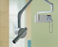
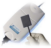
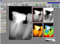
One of the basic elements of effective dental treatment is appropriate diagnosis. In our office we use the latest equipment for obtaining digital X-ray images – radiovision. The use of digital radiological technology allows to reduce the dose of radiological radiation absorbed by the patient, by up to 90% compared to the traditional method of taking pictures on X-rays. An additional advantage is that the image of the photo is obtained on the computer monitor just seconds after taking the photo – this allows you to reduce the total healing time and make computer processing photos to better illustrate important details e.g.: after duct treatment. How does a digital intraoral X-ray of the tooth look like? Here is a simplified scheme of radiological examination using radiovisiography: In common use are 2 popular methods of taking radiological images:
- simple angle method – the doctor places the radiovision sensor in a special positioner and places the positioner in the area of the teeth to be visible in the photo. The positioning of the x-ray lamp head is significantly simplified in this method.
- two-sewangle method – the doctor places a digital x-ray sensor in the area of the test tooth and asks the patient to hold it with a index finger and then position the head of the x-ray camera accordingly
With both methods, thanks to the use of digital X-ray radiovision, the photo is visible on the computer monitor as early as a few seconds after exposure and further treatment is possible.
Faqs:
- What are the advantages of digital X-ray taking? – compared to the traditional method of taking X-ray images on radiological films, radiovisiography has the same advantages, these are:
- 90% reduction of radiation dose
- reduce the wait time for a photo result to a few seconds
- computer-treated and photo archiving.
- Are there any contraindications to taking X-rays? – yes, the contraindication for X-rays is the patient's pregnancy, X-rays are also avoided in people under 18 years of age. The general principle of radiological diagnostics is to take X-ray images only if they are actually necessary.
- Is radiation harmful? – Yes, X-ray radiation is harmful, which is why it is so important to limit the doses absorbed by the patient when taking pictures as much as possible. Thanks to modern equipment, the dose absorbed during one digital X-ray is less than the dose received each day from the so-called background radiation. For example, a flight between Poland and the US results in a dose equal to dozens of digital X-rayphotos.


