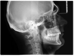
Cephalometric image – Lateral teleroentgenogram of the head in melee
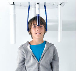
The side telerentgenogram of the head in short circuit (cephalometric photo), is the basic diagnostic photo in orthodontics. Side projection is always used before the start of orthodontic treatment due to a low dose of radiation and very high diagnostic importance. The doctor on the basis of the cephalometric photo is able to perform cephalometric analysis, which together with gypsum models and pantomography image forms the basis for the diagnosis of the bite defect. In our clinic, all X-ray images are taken in digital technology, so the radiation dose is kept to a minimum. One photo causes the adoption of radiation close to what a person receives from the environment during one day. Cephalometric photos are performed by modern Vatech PAX Green and Sirona ORTHOPHOS XG 5 cameras. The side telerentgenogram in melee (cephalometric photo) is performed mainly by orthodontic indications. A contraindication that prevents the photo from being taken is pregnancy. To take a photo, the patient is asked to remove any metal objects (earrings, chains, glasses, hairpins, hair slides). We also perform side telerentgenogram to patients outside our office. We release the result of the study digitally on a CD, pendrive media or send to the patient's email box. According to Polish law, in order to take a picture of a cephalometric x-ray, it is necessary to refer a dentist from the doctor. In the absence of such a referral, the cephalometric photo can only be taken after an earlier dental consultation during which the doctor will assess the indications for taking a side head photo.


